Have you ever wondered how a doctor can look inside the human body without making an incision?
A chest X-ray can reveal a problem in the lungs. An MRI can show detailed structures of the brain or joints. Ultrasound can show a developing fetus and even display blood flow. A CT scan can turn X-ray measurements into detailed cross-sectional views of the body.
All of these are possible because of medical imaging technology.
Medical imaging uses different technologies to obtain images and other information about the inside of the human body. These images and measurements help healthcare professionals detect disease, understand injuries, plan treatments, monitor conditions, and sometimes guide procedures. Different imaging technologies work in different ways, so each one provides a different type of information.
But medical imaging is much more than simply "taking pictures."
Why Do We Need Medical Imaging?
The human body is complex, and many problems cannot be identified by looking at the outside of the body alone.
Consider a patient who comes to a hospital with chest pain. The symptoms provide important information, but they may not be enough to determine what is happening inside the body. Imaging can provide additional information that helps answer the clinical question.
The choice of imaging depends on the clinical situation. A suspected fracture may require an X-ray, while a suspected stroke may require CT or MRI depending on the clinical circumstances. Abdominal symptoms may be investigated using ultrasound, CT, or another appropriate examination.
There is no single imaging test that is best for every situation. The modality is selected according to the clinical question, body part, information required, and patient-specific considerations.
How Does Medical Imaging Actually Create an Image?
Different imaging modalities use different forms of energy or physical phenomena to obtain information from the body.
X-ray
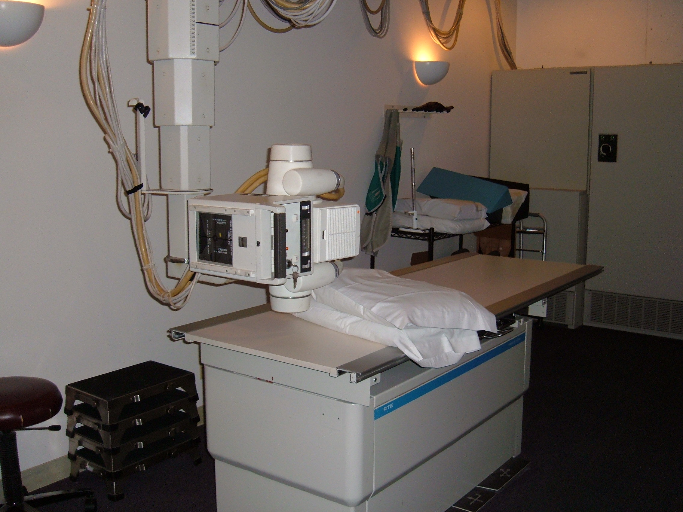
X-rays are a form of ionizing radiation. During an X-ray examination, a beam passes through the body. Different tissues attenuate, or reduce, the X-ray beam by different amounts.
The X-rays that pass through the body reach a detector, and the resulting differences are used to form the image. This is why structures such as bones and lungs appear differently on an X-ray.
Imagine shining a light through different materials. A thin transparent sheet lets most of the light through, while a thick object blocks much more of it. X-ray imaging works on a similar principle: different tissues affect the X-ray beam differently.
CT — Computed Tomography
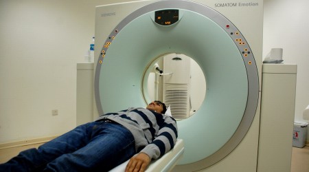
CT also uses X-rays, but instead of producing a single projection image, the X-ray source and detectors collect many measurements from different angles around the patient.
A computer then uses these measurements to reconstruct cross-sectional images, or slices, through the body.
Think about a loaf of bread. Looking at the whole loaf gives you a general idea of its shape, but cutting it into slices lets you examine what is happening inside at different levels. CT works in a similar conceptual way — although its slices are computationally reconstructed rather than physically cut.
CT can provide detailed information about bones, organs, soft tissues, and blood vessels.
MRI — Magnetic Resonance Imaging
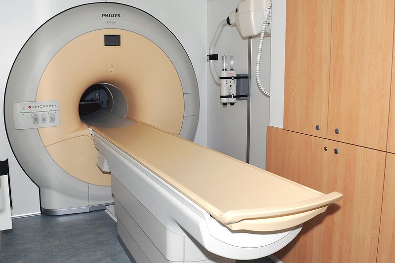
MRI works very differently from X-ray and CT. It uses a strong magnetic field and radiofrequency waves to produce signals from the body, which are then processed to create images.
MRI is particularly valuable for detailed soft-tissue imaging, including the brain, spinal cord, muscles, ligaments, and joints.
MRI does not use ionizing radiation. However, it has important safety requirements because of the strong magnetic field and radiofrequency energy involved. Like every imaging technology, MRI has its own risks, limitations, and considerations.
Ultrasound
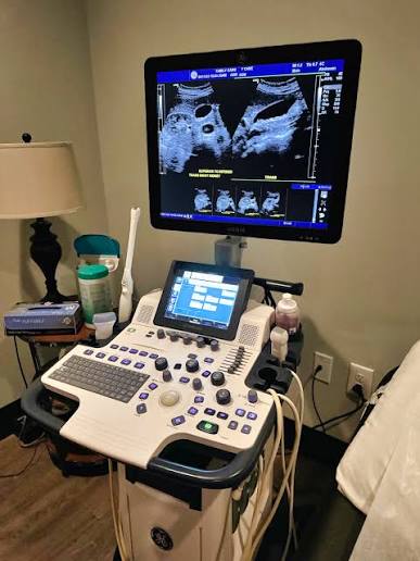
Ultrasound uses high-frequency sound waves. A transducer, or probe, sends sound waves into the body and receives the echoes that return after the waves interact with different tissues.
The ultrasound system analyzes the timing and strength of these echoes and uses the information to construct an image.
Imagine throwing a small ball toward a wall. The ball travels forward, hits the wall, and comes back. If you know how long it took to return, you can get an idea of how far away the wall is.
Ultrasound works on a similar idea. The transducer sends sound waves into the body, receives the returning echoes, and uses their timing and characteristics to build an image.
Because ultrasound can continuously send and receive sound waves, it can produce images in real time. This makes it useful for observing movement and assessing blood flow with Doppler techniques.
Nuclear Medicine
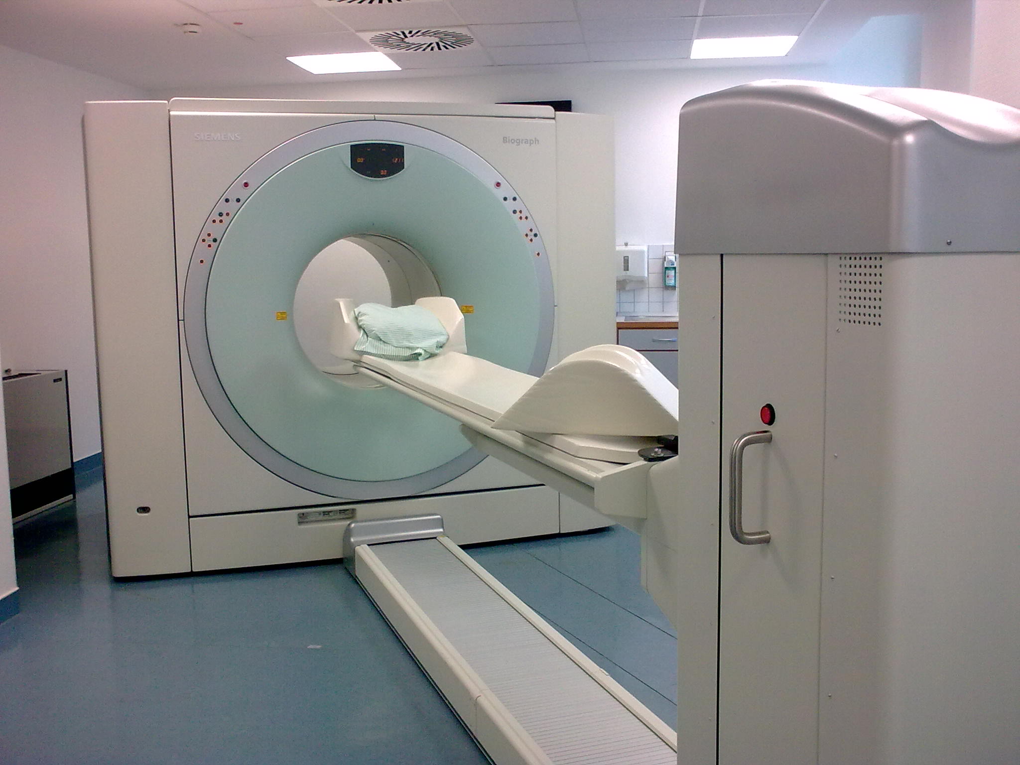
Nuclear medicine works differently from the other modalities described above. Instead of primarily sending energy into the body and measuring what comes back, a radiopharmaceutical is administered to the patient.
Depending on the examination, the radiopharmaceutical may be injected, swallowed, or inhaled. It travels to particular tissues or participates in biological processes. A detector then detects the radiation coming from the patient, and the resulting image represents the distribution of the radiopharmaceutical.
This means nuclear medicine can provide information about function and physiology, not only anatomy.
A useful way to think about the difference is: “What does this organ look like?” versus “How is this organ functioning?”
PET and SPECT are common examples of nuclear medicine imaging.
Medical Imaging Modalities at a Glance
Each modality uses a different technology and provides different types of information.
| Modality | Main Technology | Ionizing Radiation? | Commonly Good At |
|---|---|---|---|
| X-ray | X-rays | Yes | Quick projection images, especially bones and chest |
| CT | X-rays + computer reconstruction | Yes | Detailed cross-sectional anatomy |
| MRI | Magnetic field + radiofrequency waves | No | Detailed soft-tissue imaging |
| Ultrasound | High-frequency sound waves | No | Real-time imaging, soft tissues and blood flow |
| Nuclear Medicine | Radiopharmaceuticals + radiation detection | Yes | Functional and physiological information |
| Mammography | Specialized X-ray imaging | Yes | Breast imaging |
| Fluoroscopy | Continuous/rapid X-ray imaging | Yes | Real-time imaging and procedure guidance |
These modalities should not be thought of as competitors where one is simply “better” than the others. Each answers different clinical questions.
Anatomy vs Function: What Is the Difference?
One useful way to understand medical imaging is to separate structure from function.
X-rays are excellent for showing structures such as bones and providing projection images of the chest. CT provides detailed cross-sectional anatomical information. MRI is particularly useful when detailed soft-tissue information is needed. Ultrasound can show organs and movement in real time.
Nuclear medicine can go a step further by showing physiological or biochemical activity. Instead of only showing where a structure is, it can provide information about how tissue is functioning.
Hybrid imaging can combine these different types of information. For example, PET/CT combines functional information from PET with anatomical information from CT.
So medical imaging is not always simply about creating a picture. Sometimes the important question is where something is, and sometimes it is how that tissue or organ is behaving.
Does Every Imaging Modality Use Radiation?
No. This is a common misconception because the word “radiation” can sound alarming, but different imaging technologies use different forms of physical energy.
Imaging modalities that use ionizing radiation include X-ray, CT, fluoroscopy, mammography, and nuclear medicine. Ionizing radiation has enough energy to cause changes in biological tissue, so appropriate use and radiation protection are important.
MRI and ultrasound do not use ionizing radiation. MRI uses magnetic fields and radiofrequency waves, while ultrasound uses sound waves.
However, non-ionizing does not automatically mean risk-free. Each imaging technology has its own safety considerations, limitations, and appropriate uses.
What Happens During an Imaging Examination?
A medical imaging examination involves much more than pressing a button on a machine. A typical examination may involve several important steps:
- Referral / Clinical Question — Understanding why the examination is needed.
- Patient Preparation — Preparing the patient according to the requirements of the examination.
- Patient Identification and Safety Checks — Confirming patient details and relevant safety information.
- Positioning — Placing the patient correctly for the examination.
- Image Acquisition — Obtaining the required images using appropriate technical parameters.
- Image Quality Check — Ensuring that the images are suitable for their intended diagnostic purpose.
- Image Processing and Storage — Processing, storing, and transferring digital image data.
- Interpretation — The images are reviewed and interpreted by the appropriate healthcare professional.
This process requires technical knowledge, patient care, communication, safety awareness, and attention to detail.
Where Does a Medical Imaging Technologist Fit In?
The role of a medical imaging technologist is often reduced to simply operating an imaging machine. In reality, the role involves much more.
Depending on the modality and scope of practice, an imaging technologist may:
- Communicate with and prepare patients.
- Confirm examination requirements.
- Position patients correctly.
- Select appropriate technical parameters and protocols within their scope of practice.
- Operate imaging equipment.
- Produce diagnostically useful images.
- Maintain image quality.
- Apply radiation protection principles when ionizing radiation is used.
- Maintain patient safety throughout the examination.
- Work with radiologists and the wider healthcare team.
Positioning, technical factors, and equipment performance can all affect the final image. The image did not simply “appear.” There is a complete technical and clinical process behind it.
The Technology Behind the Image
Medical imaging brings together several fields, including physics, electronics, computing, engineering, and healthcare.
The imaging system first acquires information from the patient. That information is then processed using detectors or sensors, digital electronics, computers, reconstruction algorithms, and image-processing systems.
In CT, for example, the system collects measurements from multiple angles and a computer reconstructs them into cross-sectional images. Ultrasound systems process returning echoes. MRI systems process signals generated in response to radiofrequency excitation within a magnetic field.
Medical imaging is therefore a close meeting point between medicine and technology.
What About Digital Medical Images?
Modern medical images are primarily digital rather than being produced on physical film. Digital images can be processed, stored, transferred, and reviewed using computer-based systems.
Two important terms in modern medical imaging are DICOM and PACS.
DICOM is a standard used for medical imaging information and communication. PACS, or Picture Archiving and Communication Systems, are used to store and provide access to medical images within healthcare environments.
These technologies are an important part of the modern digital imaging workflow.
What Does the Future of Medical Imaging Look Like?
Medical imaging continues to evolve as scanners, computing systems, and image-processing technologies become more sophisticated.
Areas of ongoing development and research include:
- Faster and more advanced imaging systems
- Portable point-of-care ultrasound
- AI-assisted image analysis
- Quantitative imaging
- Faster image acquisition
- Improved image reconstruction
- Hybrid imaging
- Personalized imaging
- Better integration with clinical workflows
The field is gradually moving beyond simply producing pictures toward extracting more useful information from medical imaging data.
The Big Picture
Medical imaging is not one technology. It is a whole group of technologies designed to answer different clinical questions.
X-ray uses X-rays. CT uses X-rays combined with computer reconstruction. MRI uses magnetic fields and radiofrequency waves. Ultrasound uses sound waves. Nuclear medicine uses radiopharmaceuticals to provide information about biological function.
Each modality has its own physics, strengths, limitations, safety considerations, and clinical applications.
Behind every medical image is a combination of technology, physics, patient care, clinical knowledge, and human expertise.
Sources
- RadiologyInfo.org. Medical Imaging — information about diagnostic imaging procedures, technologies, radiation safety, and examinations. American College of Radiology (ACR) and Radiological Society of North America (RSNA).
- DICOM Standards Committee. Digital Imaging and Communications in Medicine (DICOM) Standard — standard for the communication and exchange of medical imaging information and associated data.
Medical imaging is not simply about creating images. It is about turning information that cannot be seen from the outside into something healthcare professionals can visualize, analyze, and use to care for a patient. That is what makes medical imaging technology such an important part of modern healthcare.