If you have studied general X-ray imaging, you already know the basic idea: X-rays pass through the body, and different tissues absorb different amounts of radiation.
But when we image the breast, we need a technique that can show very small differences in soft tissue.
This is where mammography comes in.
Mammography is a specialized type of X-ray imaging used to produce detailed images of the breast. It can help detect breast abnormalities, including some cancers, at an early stage.
So, how is mammography different from an ordinary X-ray?
Let's break it down.
What is a mammogram?
A mammogram is an X-ray image of the breast.
Mammography is the technique used to produce that image.
A mammography system is specifically designed for breast imaging. It uses a low-dose X-ray beam and a specialized detector to produce images with the detail needed to evaluate breast tissue.
Mammography can be performed as screening mammography or diagnostic mammography, depending on the reason for the examination.
How does mammography work?
The basic principle is similar to other X-ray examinations.
The X-ray tube produces X-rays and directs them toward the breast.
As the X-rays pass through the breast, different tissues absorb the radiation differently. The X-rays that pass through the breast reach the detector, which converts the information into an image.
In modern digital mammography, the detector converts the X-ray information into electrical signals that are processed into a digital image. The image can then be displayed on a computer for the radiologist to review.
But mammography has one important step that deserves a closer look:
breast compression.
Why is the breast compressed?
During mammography, the breast is gently positioned between two plates and compressed.
This can be uncomfortable, but compression is an important part of the examination.
It helps to:
- spread the breast tissue
- reduce overlapping of structures
- reduce movement and image blur
- make the breast thinner
- reduce scattered radiation
- help obtain a useful image with an appropriate radiation dose
By making the breast thinner and more evenly spread, compression helps the imaging system produce a clearer representation of the breast tissue.
What happens during a mammogram?
The examination is performed by a specially trained radiologic technologist.
The breast is placed on the imaging platform and carefully positioned. A compression paddle is then used to compress the breast.
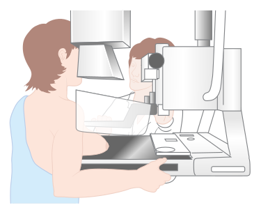
The patient is asked to remain still while the image is acquired.
Standard mammography commonly includes two main views of each breast:
- CC (craniocaudal) — a top-to-bottom view
- MLO (mediolateral oblique) — an angled view
These views provide different perspectives of the breast and help include as much breast tissue as possible.
Additional views may be obtained when needed, particularly during diagnostic examinations.
What can a mammogram show?
Mammograms can show several types of breast findings.
These may include:
- masses
- asymmetries
- architectural distortion
- calcifications
One particularly important finding is microcalcifications — very small calcium deposits that may sometimes be associated with breast cancer.
However, not every abnormal finding means cancer.
Some findings are benign, while others may require additional imaging or further evaluation.
Mammography is an important tool, but it also has limitations and does not detect every breast cancer.
Screening vs. diagnostic mammography
The terms screening and diagnostic describe why the mammogram is being performed.
Screening mammography
Screening mammography is performed when there are no signs or symptoms that have prompted the examination.
Its purpose is to look for possible abnormalities that may need further assessment.
Diagnostic mammography
Diagnostic mammography is performed when there is a specific concern.
For example, a patient may have a breast lump, nipple discharge, or a finding that needs further evaluation after a screening mammogram.
Diagnostic mammography may include additional mammographic views and, when appropriate, other imaging such as ultrasound.
Is mammography always done with film?
No.
Modern mammography is primarily digital.
In digital mammography, an electronic detector records the X-ray information and the resulting image is processed digitally.
The image can then be displayed on a workstation for evaluation and stored electronically.
You may also come across another technology called Digital Breast Tomosynthesis (DBT), often referred to as 3D mammography.
Instead of producing only a standard 2D mammographic image, DBT acquires multiple images from different angles as the X-ray tube moves over the breast.
These images are reconstructed into thin sections, which can help reduce the effect of overlapping breast tissue.
DBT is an important topic on its own, so we can explore it separately.
What about radiation dose?
Mammography uses ionizing radiation because it uses X-rays.
The examination is designed to obtain the information needed for breast evaluation while using an appropriate radiation dose.
Radiation dose in mammography depends on factors such as the breast thickness and composition, the imaging system, and the exposure settings.
This is why mammography requires carefully controlled technique and properly maintained equipment.
The goal is not simply to use the lowest possible exposure.
The goal is to produce a diagnostically useful image while keeping radiation exposure appropriately low.
Why does technique matter?
A mammogram is not simply about pressing a button and taking an image.
Positioning matters.
The breast needs to be positioned correctly so that the relevant tissue is included in the image.
Compression matters.
It helps reduce tissue overlap and motion.
Exposure factors matter.
They influence how the X-ray beam interacts with the breast and how much information reaches the detector.
Image quality matters.
A technically inadequate image may make evaluation more difficult.
This is why the role of the mammography technologist is so important. Good positioning and technique are essential parts of producing useful mammographic images.
The main idea
Mammography is a specialized form of X-ray imaging designed specifically for the breast.
The process can be simplified to:
But producing a useful mammogram involves much more than this basic sequence.
Positioning, compression, exposure, detector technology, and image quality all work together to produce an image that can be evaluated by the radiologist.
Mammography can help detect breast abnormalities, but an abnormal finding does not automatically mean cancer, and a normal mammogram does not guarantee that cancer is absent.
That is why mammography is best understood as an important part of breast imaging and evaluation, rather than a test that gives every answer by itself.
Sources
- U.S. Food and Drug Administration (FDA) — Mammography
- U.S. Food and Drug Administration (FDA) — Frequently Asked Questions About Digital Mammography
- RadiologyInfo.org — Mammography
- RadiologyInfo.org — Breast Cancer Screening