A conventional X-ray gives us a picture of the body at one particular moment. But sometimes, one picture is not enough. We may need to see what is happening while something is moving.
This is where fluoroscopy is useful.
Fluoroscopy is an X-ray imaging technique that shows a changing image on a monitor. It can be used to watch body parts, contrast material, or medical instruments while they are moving during an examination or procedure.
How does it work?
The basic idea is quite simple.
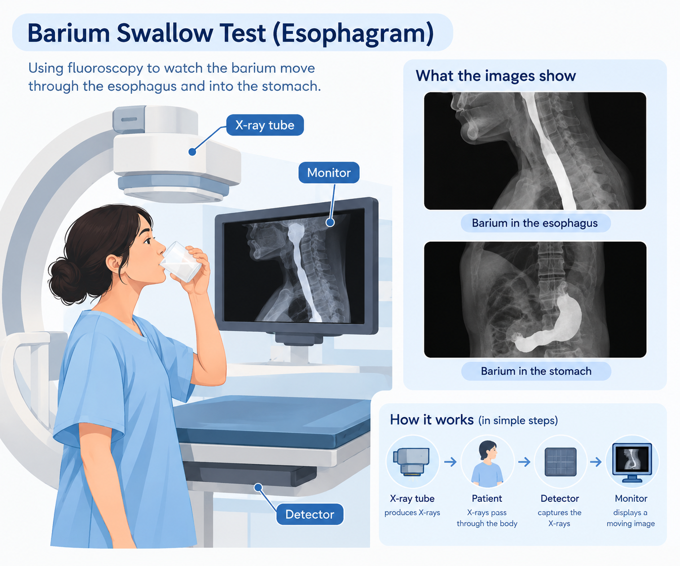
An X-ray tube produces X-rays and sends them through the part of the body being examined. Some X-rays are absorbed by the body, while others pass through and reach a detector.
In a normal X-ray, the information from the detector is used to produce a still image.
In fluoroscopy, images are produced repeatedly while the examination is taking place. The system sends these images to a monitor, where they can be viewed as a moving sequence.
So, instead of seeing just one picture, the radiologist can watch how things change from one moment to the next.
X-ray tube → Patient → Detector → Monitor
That is the basic idea behind fluoroscopic imaging.
A simple clinical example: swallowing
One of the easiest ways to understand fluoroscopy is through a swallowing examination.
Suppose a patient has difficulty swallowing. During a Video Fluoroscopic Swallowing Examination (VFSE), the patient is given different foods or liquids containing barium.
Barium helps make the material easier to see on X-ray.
As the patient swallows, fluoroscopy allows the radiologist and speech-language pathologist to watch the movement on the monitor. They can see how the material moves through the mouth and throat and assess whether the swallowing process is safe and effective.
This is something a single X-ray cannot show well.
A still image can show the position of the material.
Fluoroscopy can show how it moves.
What else can we watch with fluoroscopy?
Fluoroscopy is useful whenever movement is important to the examination or procedure.
For example, it can be used to:
- follow contrast material through the digestive system
- observe blood vessels during certain contrast examinations
- guide catheters and other instruments
- help with some orthopedic procedures
- guide the placement of devices such as stents
For example, during an upper GI examination, a patient drinks barium while the radiologist watches its movement through the esophagus, stomach, and duodenum.
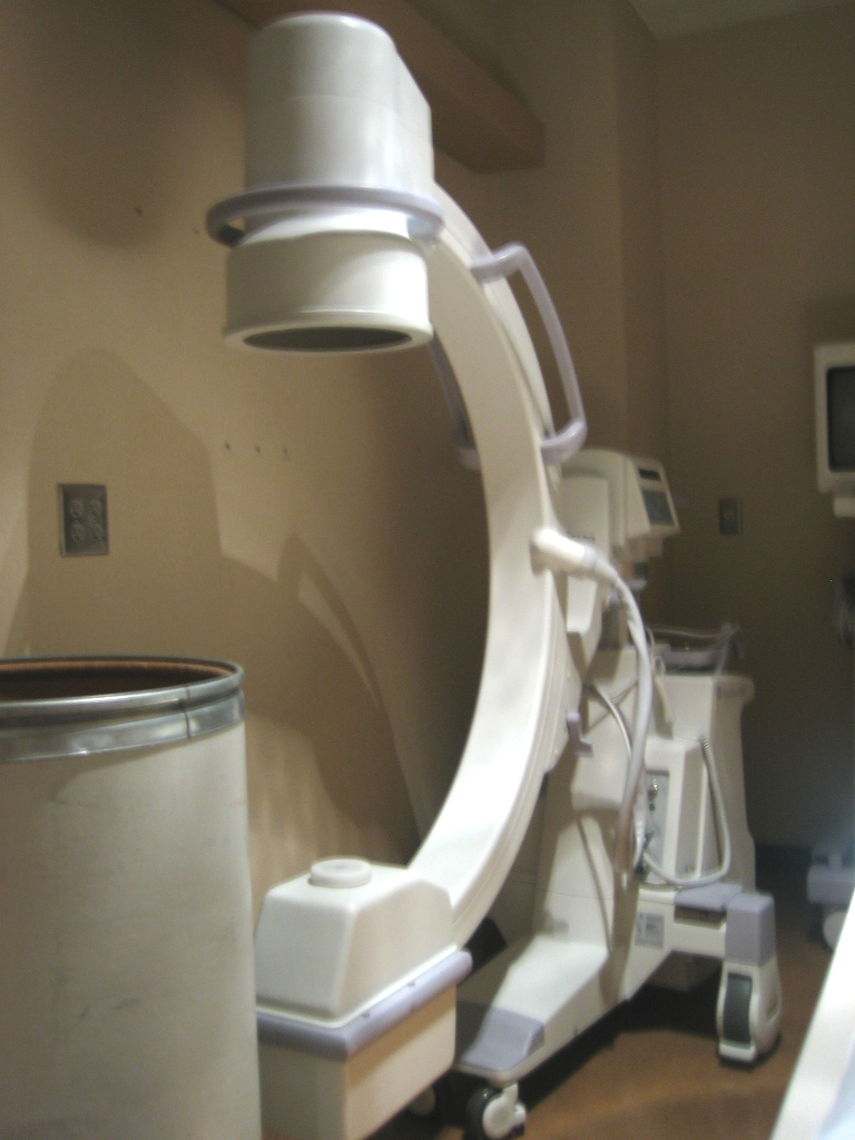
In interventional procedures, fluoroscopy can also help the physician see where an instrument is and guide it to the required location.
Is the X-ray beam always switched on?
Not necessarily.
Fluoroscopy can use a continuous X-ray beam or pulsed X-rays. With pulsed fluoroscopy, X-rays are produced in short bursts rather than continuously, while the images are still displayed as a moving sequence.
The exact settings depend on the equipment and the type of examination.
So, when we say that fluoroscopy provides a “real-time” view, it does not necessarily mean that the X-ray tube is producing radiation continuously every second.
The important point is that the system acquires images repeatedly and displays them quickly enough to follow the movement.
What about radiation dose?
Fluoroscopy uses ionizing radiation, just like conventional X-rays.
Because images may be acquired for a longer period, radiation exposure can be higher than with a single radiograph, especially during longer or more complex procedures.
This is why radiation protection is important during fluoroscopy.
The imaging team aims to use the radiation needed to obtain useful images while avoiding unnecessary exposure. Modern systems can also provide radiation-dose information and features such as last-image hold, which allows the most recent image to be reviewed without continuing the X-ray exposure.
Fluoroscopy vs. a normal X-ray
The easiest way to remember the difference is:
| Conventional X-ray | Fluoroscopy |
|---|---|
| Mainly gives a still image | Shows a moving sequence of images |
| Useful for looking at anatomy at one point in time | Useful when movement needs to be observed |
| Commonly used for chest and bone examinations | Commonly used for contrast studies and image-guided procedures |
| Usually involves a short exposure | May require repeated exposure during the examination |
Both use X-rays. The main difference is how the images are acquired and displayed.
The main idea
Fluoroscopy is useful when we need more than a picture.
It allows us to follow movement over time — whether that is contrast passing through the digestive tract, a patient swallowing, or an instrument being guided during a procedure.
So, a simple way to remember it is:
Fluoroscopy helps us see what is happening.
Sources
- U.S. Food and Drug Administration (FDA) — Fluoroscopy
- U.S. Food and Drug Administration (FDA) — Medical X-ray Imaging
- RadiologyInfo.org — Video Fluoroscopic Swallowing Exam (VFSE)
- RadiologyInfo.org — Upper GI X-ray / Barium Swallow