When you look at an ultrasound image, you may notice that simple fluid-filled structures such as the urinary bladder or a simple cyst often appear almost completely black.
At first, this can seem strange. If there is fluid inside the structure, why does the ultrasound image show almost nothing there?
The answer becomes much easier once you understand that an ultrasound image is mainly created from returning echoes.
Ultrasound Images Are Made From Echoes
An ultrasound transducer sends high-frequency sound waves into the body.
When those sound waves encounter boundaries between tissues, some of the sound is reflected back toward the transducer.
The ultrasound system uses these returning echoes to create the image.
More returning echoes → brighter appearance
Fewer returning echoes → darker appearance
So when a structure appears black, it does not mean that there is nothing there.
It means that very little ultrasound energy is returning from that region to produce image brightness.
When a structure produces essentially no internal echoes, it is described as anechoic.
Anechoic structures therefore appear black on the ultrasound image.
So, What Is Different About Simple Fluid?
Many solid tissues contain small structures and interfaces that can scatter or reflect ultrasound in different directions.
These interactions create returning echoes, which contribute to the gray-scale appearance of the image.
Simple fluid is different.
A simple fluid-filled space is relatively uniform and has very few internal structures that produce significant echoes.
As a result, very little sound is scattered back from inside the fluid toward the transducer.
Few internal echoes → dark / anechoic appearance
This is the main reason simple fluid often looks completely black on ultrasound.
But Is the Ultrasound Sound Actually Stopping There?
No.
This is one of the most important points to understand.
The fact that fluid looks black does not mean that the ultrasound beam cannot travel through it.
In fact, ultrasound generally travels through simple fluid quite well.
There is an important difference between:
Sound travelling through the fluid
and
Sound returning from inside the fluid.
Simple fluid produces very few internal echoes, so it looks black. But the ultrasound beam can continue travelling through the fluid.
What Happens When the Sound Reaches the Other Side?
Now we get to the interesting part.
Imagine an ultrasound beam entering a simple cyst.
Inside the cyst, there are very few structures to reflect or scatter the sound.
Therefore:
↓
Very few internal echoes are produced
↓
Very little sound returns from inside the cyst
↓
The cyst appears black
↓
Sound continues through the fluid
↓
The beam reaches the tissue behind the cyst
↓
More sound reaches the deeper tissue
↓
Stronger returning echoes may be produced
↓
The region behind the cyst may appear brighter
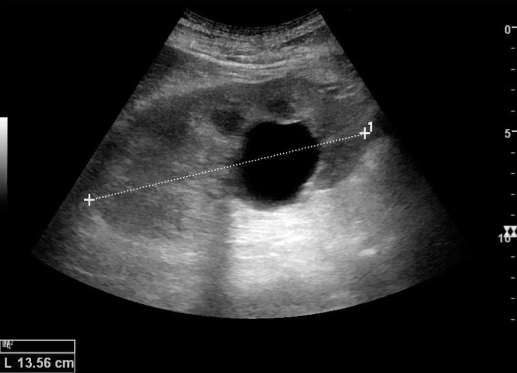
This increased brightness behind a fluid-filled structure is called posterior acoustic enhancement.
Why Does Fluid Allow More Sound Through?
To understand this, we need one important ultrasound concept: attenuation.
Attenuation means that the energy of the ultrasound beam decreases as it travels through tissue.
Different tissues attenuate ultrasound by different amounts.
Simple fluid generally causes relatively little attenuation compared with many soft tissues.
This means that more of the ultrasound beam can reach structures deeper than the fluid-filled space.
Less attenuation → more sound reaches deeper tissue
Because more sound reaches the tissue behind the fluid, the returning echoes from that deeper tissue can be stronger than expected.
The deeper region may therefore appear brighter.
A Simple Cyst Is a Good Example
A simple cyst is one of the clearest examples of this phenomenon.
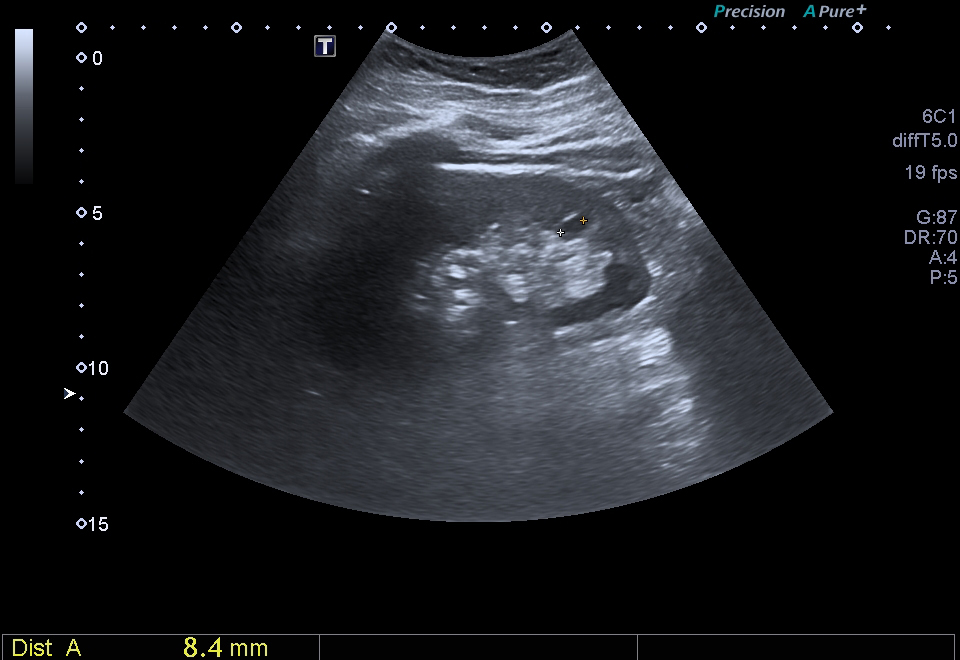
On ultrasound, a typical simple cyst commonly has:
- Well-defined margins
- Anechoic interior
- No internal echoes
- Posterior acoustic enhancement
The combination of a black interior and increased brightness behind the cyst is therefore a useful ultrasound finding.
What About the Urinary Bladder?
The urinary bladder provides another familiar example.
When the bladder contains relatively simple urine, the urine can appear anechoic, producing a dark or black appearance.
The ultrasound beam can continue through the urine and reach tissues deeper to the bladder.
As a result, the tissues behind the bladder may appear relatively brighter.
Again, the black appearance does not mean that the ultrasound beam has stopped.
Does All Fluid Look Completely Black?
No.
The classic black appearance mainly describes simple fluid.
Fluid can contain things such as:
- Blood
- Protein
- Pus
- Cells
- Debris
These components can create internal echoes.
Therefore, a fluid collection may appear gray or have a more complex internal appearance instead of being completely anechoic.
Simple fluid → usually anechoic
Complex fluid → may contain internal echoes
Black Does Not Always Mean Fluid
Another important point is that a black area on an ultrasound image does not automatically mean that it contains fluid.
Different ultrasound phenomena can produce dark appearances for different reasons.
For example, acoustic shadowing behind a highly attenuating or strongly reflecting structure, such as a stone, can produce a dark region.
That dark region is produced by a different mechanism from the anechoic appearance of simple fluid.
So when looking at an ultrasound image, it is better to ask:
rather than automatically assuming:
Anechoic vs Hypoechoic
These two terms are commonly used when describing ultrasound appearance.
Anechoic means that essentially no internal echoes are detected from the structure.
It therefore appears black.
Hypoechoic means that a structure produces fewer echoes than the surrounding tissue.
A hypoechoic structure can therefore look darker than its surroundings, but it may still contain visible gray-level echoes.
Anechoic → essentially no internal echoes → black
Hypoechoic → fewer echoes than surrounding tissue → darker gray
The Interesting Part: Black Inside, Bright Behind
This is probably the most useful concept to take away from this article.
↓
Very few internal structures produce echoes
↓
Very little sound returns
↓
Cyst appears black
↓
Sound continues through the fluid with relatively little attenuation
↓
More sound reaches the tissue behind
↓
Stronger echoes return
↓
Area behind may appear brighter
That last part is called posterior acoustic enhancement.
Once you understand this, the black appearance of simple fluid becomes much less mysterious.
The Whole Idea in One Simple Example
Imagine a simple cyst inside soft tissue.
The cyst appears black because there are very few internal echoes.
But the sound does not simply disappear at the cyst.
It continues travelling through the fluid, which generally has relatively low attenuation.
This allows more sound to reach the tissue behind the cyst.
The deeper tissue can therefore produce stronger returning echoes, making the region behind the cyst appear brighter.
Black inside → very few internal echoes
Bright behind → more sound reaches deeper tissue
The image is therefore showing us something about how ultrasound interacts with the structure, not simply whether the structure contains something or not.
Quick Recap
- Ultrasound images are created mainly from returning echoes.
- Simple fluid produces very few internal echoes.
- Very few echoes produce a dark or anechoic appearance.
- The ultrasound beam can still travel through the fluid.
- Simple fluid generally causes relatively low attenuation.
- More sound can therefore reach the tissue behind the fluid.
- The deeper tissue may appear brighter.
- This increased brightness is called posterior acoustic enhancement.
- Not every fluid collection is completely black.
- A black area does not automatically mean that it is fluid.
The Key Idea
Simple fluid looks black on ultrasound because it produces very few internal returning echoes.
The ultrasound beam is still able to travel through the fluid.
Because simple fluid generally causes relatively little attenuation, more sound can reach deeper tissue, which can make the region behind the fluid appear brighter.
Simple fluid → few echoes → black appearance
Low attenuation → more sound reaches deeper tissue → posterior acoustic enhancement
Once you understand this relationship, the appearance of simple fluid on ultrasound becomes something you can reason through rather than simply memorize.
Takeaway
When simple fluid looks black on an ultrasound image, it does not mean that there is “nothing” inside.
It means that the fluid produces very few internal echoes, so little sound returns to the transducer from inside the fluid.
At the same time, the ultrasound beam can continue through the fluid and reach deeper tissue.
That combination explains why a simple fluid-filled structure can look black inside but brighter behind.
Sources & Further Reading
-
NCBI / PMC
— Clinical Ultrasound Physics
Basic ultrasound physics, echoes, attenuation and image formation.
Read the article -
NCBI Bookshelf / StatPearls
— Ultrasound Physics and Instrumentation
Ultrasound wave propagation, attenuation, reflection and image formation.
Read the chapter -
Society of Radiologists in Ultrasound Consensus
— Consensus terminology and characterization of cystic lesions
Ultrasound appearance and terminology related to simple and complex cystic structures.
Read the article