Ultrasound images are created from the echoes produced when sound waves travel through the body. What appears behind a structure depends partly on what happens to the ultrasound beam as it passes through it.
Two important appearances are acoustic shadowing and posterior acoustic enhancement. They are almost opposite in appearance: one makes the region behind a structure look darker, while the other makes it look relatively brighter.
First, what does “posterior” mean?
In ultrasound, posterior simply means the area deeper than the structure being examined.
So when we talk about a posterior artifact, we are describing an appearance that occurs behind the structure along the path of the ultrasound beam.
Acoustic Shadowing
Acoustic shadowing occurs when the ultrasound beam is greatly reduced as it encounters a strong reflector, highly attenuating structure, or another structure that limits transmission of sound.
Because much less sound reaches the tissues deeper to it, fewer returning echoes are available from that region. The area behind the structure therefore appears darker than the surrounding tissue.
What can cause acoustic shadowing?
Common causes include structures that strongly reflect, scatter, or attenuate the ultrasound beam, such as:
- Gallstones and other stones
- Calcifications
- Bone
- Gas or air-filled bowel
- Some highly attenuating tissues or lesions
The exact appearance of the shadow depends on the structure and the way the ultrasound beam interacts with it.
A classic example: a stone
A stone can strongly reflect and attenuate the ultrasound beam. As a result, relatively little sound reaches the tissues behind it.
On the image, this produces a dark region extending deep to the stone. This is called posterior acoustic shadowing.
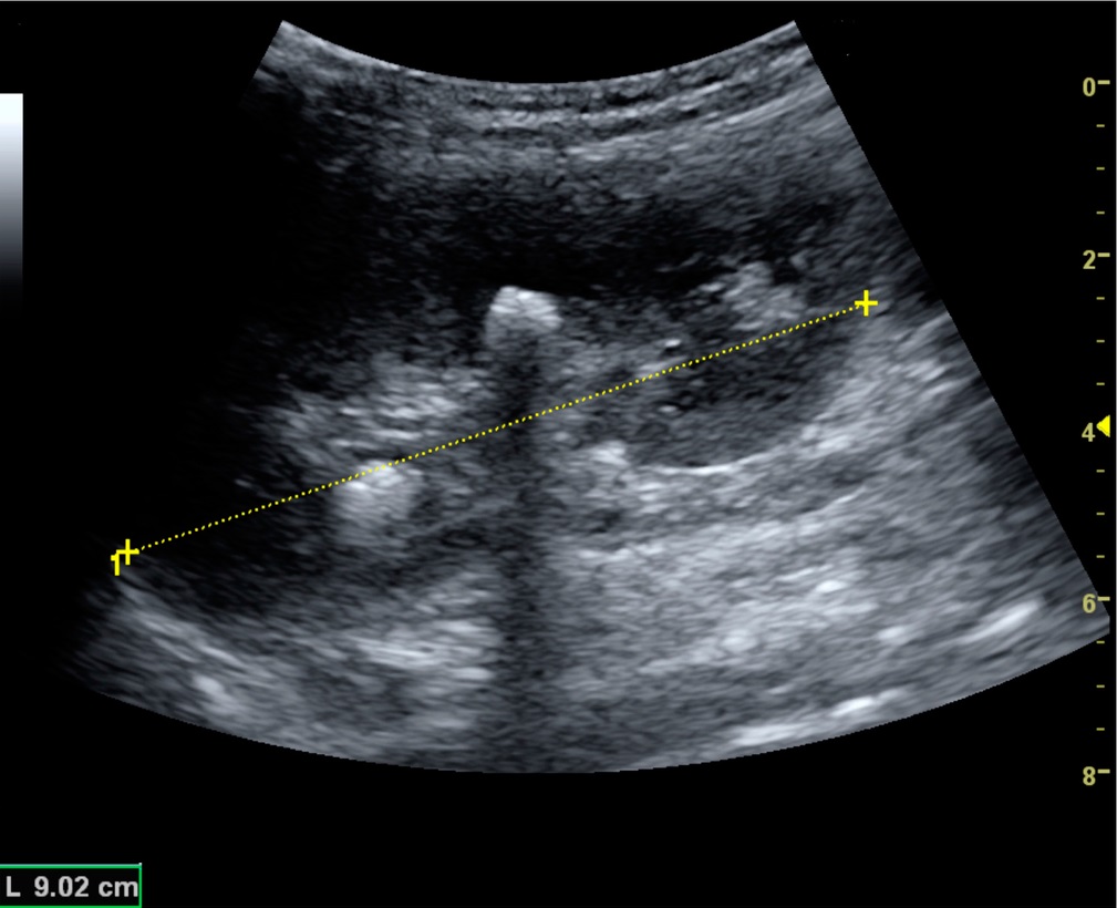
Clean shadowing vs dirty shadowing
Not all acoustic shadows look exactly the same.
Clean shadowing
A clean shadow is usually a well-defined dark region behind a strongly attenuating or reflecting structure. Stones and calcifications are common examples.
Dirty shadowing
A dirty shadow has a less sharply defined appearance and may contain internal echoes or reverberation artifacts. It is commonly associated with gas or air.
Posterior Acoustic Enhancement
Posterior acoustic enhancement is essentially the opposite visual effect.
It occurs when the ultrasound beam passes through a structure that attenuates sound relatively little. More sound can therefore reach the tissues deeper to that structure and return to the transducer.
The deeper region may consequently appear brighter than the surrounding tissue.
A classic example: a simple cyst
A simple cyst contains fluid and usually has very few internal structures to produce echoes. The fluid therefore appears anechoic, or black, on the ultrasound image.
At the same time, sound travels through the fluid with relatively little attenuation compared with surrounding soft tissue. More sound reaches the tissues behind the cyst, producing increased brightness there.
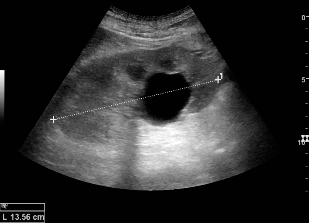
Why is the cyst itself black but the area behind it brighter?
These two appearances are caused by different parts of the same ultrasound interaction.
Inside a simple cyst, there are few structures to reflect sound back to the transducer, so the cyst appears dark.
However, the sound beam is not simply “stopped” by the cyst. Because the fluid causes relatively little attenuation, more sound continues through the cyst and reaches the tissue behind it.
That increased transmission contributes to the brighter appearance deep to the cyst.
Shadowing vs Enhancement
| Feature | Acoustic Shadowing | Posterior Enhancement |
|---|---|---|
| Appearance behind structure | Darker | Brighter |
| What happens to the beam? | Transmission is greatly reduced | Relatively more sound is transmitted |
| Effect on deeper echoes | Fewer or weaker returning echoes | More returning echoes |
| Common example | Stone or calcification | Simple fluid-filled cyst |
Why does enhancement look brighter?
Ultrasound systems compensate for the expected decrease in echo strength as sound travels deeper into the body. This is part of time-gain compensation (TGC).
When the beam passes through a structure with relatively low attenuation, such as simple fluid, more sound than expected may reach the deeper tissues. The resulting echoes can therefore appear relatively brighter.
So posterior enhancement is not simply because the tissue behind the cyst has suddenly become more reflective. It is related to the way the ultrasound beam was transmitted through the structure before reaching that tissue.
What about the edges of a cyst?
You may sometimes notice a thin dark shadow along the lateral edges of a rounded cyst. This is called edge shadowing.
It can occur because the curved boundary of the cyst changes the direction of the ultrasound beam through refraction. Some sound is redirected away from the transducer, leaving less returning signal in that region.
This is different from the broad posterior shadow produced by a strongly attenuating structure such as a stone.
Are these really “artifacts”?
Yes. In ultrasound, an artifact is an image appearance that does not represent the anatomy exactly as it exists physically.
But that does not mean artifacts are useless or simply errors to ignore. Recognizing characteristic artifacts can provide useful information about how the ultrasound beam interacted with a structure.
Why are these appearances useful?
Shadowing and enhancement can help you understand what you are seeing on an ultrasound image.
For example, a dark region behind a gallstone can support the identification of the stone, while posterior enhancement behind a simple cyst is a common feature of fluid-filled structures.
However, these appearances should not be interpreted on their own. Enhancement and shadowing are not diagnoses and are not specific to only one type of lesion. Other sonographic features and the clinical context are considered during interpretation.
A better way to remember them
Instead of memorizing two separate definitions, ask one question:
If much less sound reaches the deeper tissues, the region behind the structure tends to look darker: shadowing.
If relatively more sound reaches the deeper tissues, the region behind the structure may look brighter: posterior enhancement.
Quick recap
- Acoustic shadowing: reduced transmission of sound leads to reduced echoes from deeper tissues, producing a darker region behind the structure.
- Posterior acoustic enhancement: relatively low attenuation allows more sound to reach deeper tissues, producing increased brightness behind the structure.
- Clean shadowing is commonly associated with stones or calcifications.
- Dirty shadowing is commonly associated with gas or air.
- Edge shadowing can occur at the curved edges of cystic structures because of refraction.
- These appearances are useful clues, but they should be interpreted together with the rest of the ultrasound examination.
Key Takeaway
The easiest way to understand these two ultrasound appearances is to follow the beam.
Shadowing: less sound gets through → deeper echoes are reduced → the area behind looks darker.
Enhancement: relatively more sound gets through → deeper echoes are stronger → the area behind looks brighter.
Once you understand what happened to the ultrasound beam, the appearance becomes much easier to remember.
Sources & Further Reading
- Clinical Significance of US Artifacts — Radiographics / RSNA
- Intensive Care Ultrasound: I. Physics, Equipment, and Image Quality
- Practical and illustrated summary of updated BI-RADS for ultrasonography
- Ultrasonography of the Kidney: A Pictorial Review
- Ultrasound Physics and Technical Facts for the Beginner — SEMPA Sonoguide